Cell Structure:
Candidates should be able to:
Describe, interpret
electron micrographs,
rough and smooth endoplasmic reticulum, Golgi body,
mitochondria, chloroplasts,
ribosomes, lysosomes,
cell surface membrane,
nuclear envelope, centrioles, nucleus and nucleolus.
Candidates should be able to:
Outline
functions of the membrane systems and organelles
PROKARYOTES ( pro, before; karyon, nucleus ) lack true nuclei ie. their genetic material (DNA) is not enclosed by nuclear membranes, and lies free in the cytoplasm.
eg. Bacteria.
EUKARYOTES ( eu, true ) are more complex and are characterised by a true nucleus with their genetic material being enclosed by the nuclear envelope.
eg. Protoctists, fungi, green plants, animals.
Are you able to tell the differences?


Are you able to identify the different cell Organelles?

Cell Parts:
NUCLEUS
Found in eukaryotic cells.
Largest cell organelle
Controls cell activities
NUCLEAR ENVELOPE
NUCLEAR PORES
PERINUCLEAR SPACE
NUCLEOPLASM
CHROMATIN – DNA, HISTONES
CHROMOSOMES
HETEROCHROMATIN
EUCHROMATIN
NUCLEOLUS
CONTROLS CELL ACTIVITIES
INVOLVED IN CELL DIVISION
PROTEIN SYNTHESIS
NUCLEOLUS MANUFACTURES RIBOSOMES
WHICH ARE THE CELL ORGANELLES
RELATED THROUGH THE
ENDOMEMBRANE SYSTEM?
Rough endoplasmic reticulum
Smooth endoplasmic reticulum
Golgi apparatus
Consists of flattened, membrane-bound sacs called cisternae.
Complex system of membranes running through the cytoplasm of all eukaryotic cells.
Continuous with the nuclear membrane.
2 types :
ROUGH ENDOPLASMIC RETICULUM
SMOOTH ENDOPLASMIC RETICULUM
STRUCTURE OF ENDOPLASMIC RETICULUM
ROUGH ER
Covered with ribosomes
Sheet-like
SMOOTH ER
No ribosomes
More tubular
RIBOSOMES
RIBOSOMES
Consists of a small subunit and a large subunit
Ribosomes are found freely floating in the cytosol or attached to the endoplasmic reticulum
Site of protein synthesis
MICROSOMES
Small membrane-bound sacs.
Formed during the homogenisation procedure.
Rough ER is broken into small pieces and they reseal into vesicles.
ie. Microsomes
Microsomes do not exist as such in intact cells
FUNCTIONS OF ENDOPLASMIC RETICULUM
ROUGH ER
Transport of proteins -synthesised at the ribosomes on its surface
Synthesise phospholipids
SMOOTH ER
Lipid synthesis
Detoxification of drugs and poisons
Carbohydrate synthesis
Store calcium
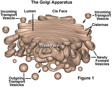
GOLGI APPARATUS
STRUCTURE:
Cisternae - stack of flattened, membrane-bound sacs
Golgi vesicles.
‘cis’ face – new cisternae formed
‘trans’ face – cisternae break up into vesicles

FUNCTIONS OF GOLGI APPARATUS
Transport and chemically modify the materials contained within it.
ve.g. glycoproteins
Sorts and targets completed materials to different parts of the cell/ for secretion out of the cell.
Formation of lysosomes
Vesicles budded off from the Golgi apparatus fuse with the plasma membrane
replace membrane
LYSOSOMES
LYSIS : SPLITTING
SOMA : BODY
LYSOSOMES
WHAT IS THE DIFFERENCE BETWEEN LYSOSOMES AND PEROXISOMES ?
LYSOSOMES AND PEROXISOMES
STRUCTURE OF LYSOSOMES
USUALLY SPHERICAL SACS BOUNDED BY A SINGLE MEMBRANE
0.2 – 0.5 um in diameter
CONTAINS HYDROLYTIC ENZYMES
CONTENTS ARE ACIDIC AND THE ENZYMES HAVE A LOW OPTIMUM PH
STRUCTURE OF PEROXISOMES
Spherical
0.3 – 1.5 um in diameter
Bounded by single membrane
Derived from the ER
Presence of the enzyme catalase which catalyses the decomposition of H2O2 to water and oxygen.
Eg. Glyoxysomes, leaf peroxisome, non-specialised peroxisomes.
LOW PH IN LYSOSOMES
TYPES OF LYSOSOMES
PRIMARY LYSOSOMES
Enzymes are synthesised on rough ER and transported to the Golgi Apparatus. Golgi vesicles containing the processed enzymes later bud off and are called primary lysosomes.
SECONDARY LYSOSOMES
Primary lysosomes may fuse with vacuoles formed by endocytosis ( infolding of the plasma membrane ) to form secondary lysosomes.
FUNCTIONS OF LYSOSOMES
DIGESTION OF MATERIALS
AUTOPHAGY
RELEASE OF ENZYMES OUTSIDE THE CELL
AUTOLYSIS
ELECTRON MICROGRAPH OF MITOCHONDRIA
Muscle Cell Mitochondrion (TEM x190,920). This image is copyright Dennis Kunkel at www.DennisKunkel.com, used with permission.
STRUCTURE OF MITOCHONDRIA
VARIABLE SHAPE AND SIZE
BOUNDED BY TWO MEMBRANES
INTERMEMBRANE SPACE
SMOOTH OUTER UNIT MEMBRANE
INNER MEMBRANE THROWN INTO FOLDS – CRISTAE
CRISTAE – ELEMENTARY PARTICLES (with head piece, stalk and base)
FLUID-FILLED INTRACRISTAL SPACE
MATRIX – ribosomes, circular DNA, enzymes
FUNCTION OF MITOCHONDRIA
SITE OF AEROBIC RESPIRATION
PRODUCTION OF ATP
MATRIX – KREBS’ CYCLE
CRISTAE – ELECTRON TRANSPORT
What happens to old, worn-out mitochondria?
CAN THE MITOCHONDRIA DIVIDE ?
MITOCHONDRIAL DNA
ORIGIN OF MITOCHONDRIA
WHERE DO MITOCHONDRIA COME FROM ?
MITOCHONDRIA AND ENDOSYMBIOSIS
During the 1980s, Lynn Margulis proposed the theory of endosymbiosis to explain the origin of mitochondria and chloroplasts from permanent resident prokaryotes. According to this idea, a larger prokaryote (or perhaps early eukaryote) engulfed or surrounded a smaller prokaryote some 1.5 billion to 700 million years ago.
The basic events in endosymbiosis. Image from Purves et al., Life: The Science of Biology, 4th Edition, by Sinauer Associates (www.sinauer.com) and WH Freeman (www.whfreeman.com), used with permission.
Instead of digesting the smaller organisms the large one and the smaller one entered into a type of symbiosis known as mutualism, wherein both organisms benefit and neither is harmed. The larger organism gained excess ATP provided by the "protomitochondrion" and excess sugar provided by the "protochloroplast", while providing a stable environment and the raw materials the endosymbionts required. This is so strong that now eukaryotic cells cannot survive without mitochondria (likewise photosynthetic eukaryotes cannot survive without chloroplasts), and the endosymbionts can not survive outside their hosts. Nearly all eukaryotes have mitochondria. Mitochondrial division is remarkably similar to the prokaryotic methods that will be studied later in this course.
MITOCHONDRIAL INHERITANCE
WHAT HAVE YOU READ ABOUT MITOCHONDRIAL INHERITANCE ?
PLANT CELLS UNDER LIGHT MICROSCOPE
STRUCTURE OF CHLOROPLAST
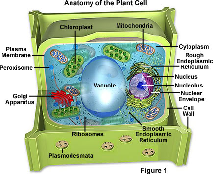
ELECTRON MICROGRAPH OF PLANT CELL
ELECTRON MICROGRAPH OF CHLOROPLASTS
ELECTRON MICROGRAPH OF CHLOROPLAST
STRUCTURE OF CHLOROPLAST
BICONVEX IN SECTION AND CIRCULAR IN SURFACE VIEW
TWO MEMBRANES – CHLOROPLAST ENVELOPE
INTERMEMBRANE SPACE
THYLAKOIDS/LAMELLAE
STROMA – circular DNA, ribosomes, enzymes, starch grains, lipid globules
FUNCTION OF CHLOROPLAST
Chloroplast provide sites on which the biochemical and photochemical reactions can occur without interference from those going in the rest of the cytoplasm.
Membrane system : site of light reaction
Stroma : site of dark reaction
STRUCTURE OF CHLOROPLAST
WHICH ARE THE ORGANELLES
THAT FORM THE CELL’S INTERNAL
SKELETON / OR ARE RELATED TO
MOVEMENT?
CENTRIOLES
A pair of cylindrical, rod-like structures
Long axes of centrioles are at 900 degrees to one another
Each centriole contains nine triplets of microtubules arranged in a ring
Found in region known as centrosome
Located close to the nucleus
During cell division, centrioles replicate and move to opposite ends of the cell.
Cells of higher plants lack centrioles
CENTRIOLES
FUNCTION :
Play a role in nuclear division in animal cells.
Centrosome divides and a pair of centrioles move to opposite poles of the cell where they help to organise the formation of spindle fibres.
IMPLICATION !
CELLS OF HIGHER PLANTS LACK CENTRIOLES
WHAT HAPPENS DURING NUCLEAR DIVISION ?
Microtubule-organising centers
Pericentriolar material surrounding the centrioles in the centrosome
Contains ring-shaped structures composed of tubulin
Can nucleate the assembly of microtubules in animal cells
Centrosome of plants and fungi lack centrioles but still contain microtubule-organising centers.
MOTILE CELLS
Centrioles divide to produce basal bodies from which flagella and cilia develop.
Cilia and flagella contain a characteristic “ 9 + 2 “ arrangement of microtubules.
CENTRIOLES VS CILIA
CYTOSKELETON
Network of protein fibres
Provides structural support
Controls cell movement
Provides anchorage for organelles and directs their movement within the cell.
CYTOSKELETON
Consists of:
Microtubules
Microfilaments
Intermediate filaments
WHAT IS THE OTHER
NON-MEMBRANOUS
CYTOPLASMIC INCLUSION ?
RIBOSOMES
STRUCTURE :
Consists of 2 subunits
Made of of ribosomal RNA and protein
FUNCTION :
Site of protein synthesis
ER-bound ribosomes make proteins which are secreted at the cell surface
Free ribosomes make proteins for use inside the cell
RIBOSOMES
TYPES:
70S – found in prokaryotes
80S – found in eukaryotes
2 populations
Free ribosomes
ER-bound ribosomes
ER-BOUND RIBOSOMES
POLYSOMES
Checkpoint:
What is the site of enzyme synthesis in cells?
Golgi apparatus
Lysosomes
Ribosome
Smooth endoplasmic reticulum
(J95 Q2)
WHAT IS THE CYTOPLASMIC
GROUND SUBSTANCE ?
CYTOPLASMIC GROUND SUBSTANCE
Aqueous ground substance containing
cell organelles and other inclusions.
CYTOSOL:
soluble part of the cytoplasm
90% water
Forms a solution which contains all the fundamental biochemicals of life.
Site of metabolic pathways
‘CYTOPLASMIC STREAMING’ – active mass movement of cytoplasm
WHAT ARE THE STRUCTURES CHARACTERISTIC OF
PLANT CELLS ?
FEATURE UNIQUE TO PLANT CELLS
CHLOROPLASTS
CELL WALL
VACUOLE
CELL WALL
TYPES :
PRIMARY WALL – cellulose microfibrils, matrix
SECONDARY WALL – extra layers of cellulose, lignin
SURFACE VIEW OF CELL WALL SHOWING CELLULOSE MICROFIBRILS
CELL WALL
FUNCTIONS :
Mechanical strength and skeletal support
Allows development of turgidity
Limits and controls cell growth and shape
Cuticle reduces water loss and risk of infection
PLASMODESMATA
PLASMODESMATA
VACUOLES
STRUCTURE :
large central vacuole
surrounded by tonoplast
cell sap
FUNCTIONS :
osmotic uptake of water
Contains pigments, hydrolytic enzymes, waste products, food reserves
HOW ARE THE EUKARYOTIC ORGANELLES CLASSIFIED UNDER FOUR FUNCTIONAL CATEGORIES ?
EUKARYOTIC ORGANELLES AND THEIR FUNCTIONS


1 comments:
Thanks for sharing these worthy information with us. I liked it. But don't forget to share further informative and similar kinds of post about Plant and Animal cells. Keep up the good work. Happy blogging!
Post a Comment